May-Thurner syndrome
Compression of the left common iliac vein between the spine and the right common iliac artery, which crosses in front of it. This persistent compression can narrow the vein, impede blood return from the left leg and promote thrombosis.
Is it common?
The anatomical compression is common: in people without symptoms, compression of more than 50% has been described in about 1 in 4. But only some develop problems. That is why we speak of a syndrome when the compression is accompanied by symptoms or thrombosis.
Forms of presentation
Without thrombosis (non-thrombotic lesion)
- Venous claudication: congestion, heaviness and pain in the left leg that increase when walking and force you to stop.
- Swelling of the left leg.
- Varicose veins, including pelvic, vulval or groin varicose veins, and symptoms of pelvic congestion.
- In advanced cases, skin changes and ulcers.
With thrombosis
May-Thurner syndrome is an important cause of left iliofemoral deep vein thrombosis, particularly in young women, sometimes triggered by the contraceptive pill, pregnancy or immobilisation. It is an extensive thrombosis that requires urgent treatment.
Diagnosis
Duplex ultrasound points to the diagnosis, and MRI or CT shows the anatomy. Confirmation and precise measurement of the narrowing are obtained with venography and intravascular ultrasound (IVUS), which is more sensitive than venography alone for these lesions.
Treatment
- No symptoms: it is not treated simply because of a finding on a scan.
- Mild symptoms: conservative measures and compression stockings.
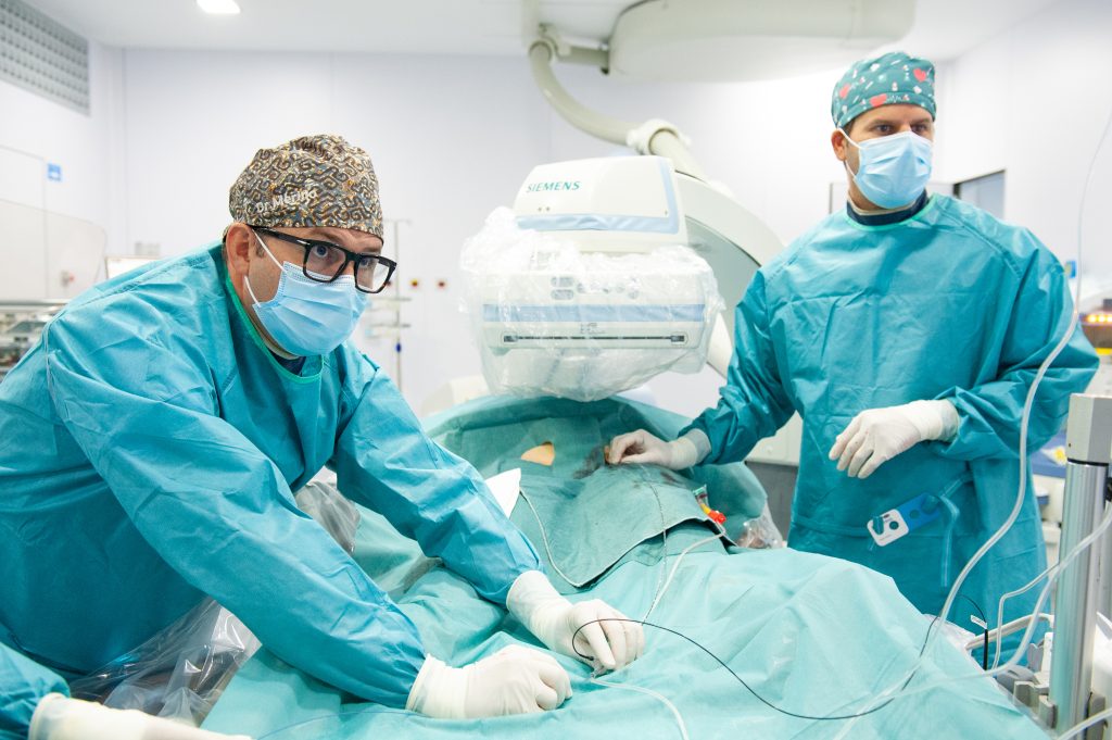
- Significant symptoms or thrombosis: endovascular treatment. Through a vein in the groin or behind the knee, I confirm the lesion with IVUS and place a venous stent that keeps the vein open at its normal calibre. If there is a recent clot, it can first be removed using thrombectomy techniques.
After stenting, antithrombotic treatment is prescribed for a period and follow-up is carried out with duplex ultrasound. The 2022 European guidelines also consider surgery or hybrid techniques for complex obstructions of the iliac veins with resistant ulcers, severe post-thrombotic syndrome or disabling venous claudication.
Results of venous stenting
A meta-analysis of 37 studies with 2,869 patients analysed stenting of iliac vein obstructions. At one year, the stents remained open without further intervention in:
- 96% of compressions without thrombosis, such as non-thrombotic May-Thurner syndrome.
- 87% after acute thrombosis.
- 79% of chronic post-thrombotic obstructions (94% including those that needed a touch-up procedure).
Technical success was achieved in 94–96% of cases, with few serious complications: major bleeding in 1.1% or less and pulmonary embolism in 0.9% or less. At Clínica Rotger I measure the compression and choose the stent using intravascular ultrasound (IVUS).
Link with pelvic congestion
When the left iliac vein is compressed, blood from the leg and pelvis seeks alternative routes and the pelvic veins dilate. That is why May-Thurner syndrome can cause vulval, groin or inner-thigh varicose veins and symptoms of pelvic congestion. In these cases, treating the compression is an essential part of treatment.
Frequently asked questions
Why does it almost always affect the left leg?
Because it is the left iliac vein that gets trapped between the right iliac artery, which crosses in front of it, and the spine.
Is the stent permanent?
Yes, the venous stent remains in place permanently. Afterwards, follow-up with duplex ultrasound checks that it stays open.
Will I need to take anticoagulants?
After stenting, antithrombotic treatment is prescribed for a period. How long depends mainly on whether there has been a previous thrombosis.
An iliac vein compression was seen on my CT scan. Do I need a stent?
Not necessarily. Around one in four people without symptoms has a compression of more than 50%. It is only treated when it explains the symptoms or has caused a thrombosis.
Do you see patients from outside Mallorca?
Yes. You can send me your test results: I review them in advance and we hold a first video consultation. Here is how it works.
References
- Kibbe MR, Ujiki M, Goodwin AL, Eskandari M, Yao J, Matsumura J. Iliac vein compression in an asymptomatic patient population. J Vasc Surg. 2004;39(5):937-943.
- Gagne PJ, Tahara RW, Fastabend CP, et al. Venography versus intravascular ultrasound for diagnosing and treating iliofemoral vein obstruction. J Vasc Surg Venous Lymphat Disord. 2017;5(5):678-687.
- De Maeseneer MG, Kakkos SK, Aherne T, et al. European Society for Vascular Surgery (ESVS) 2022 Clinical Practice Guidelines on the Management of Chronic Venous Disease of the Lower Limbs. Eur J Vasc Endovasc Surg. 2022;63(2):184-267.
- Kakkos SK, Gohel M, Baekgaard N, et al. European Society for Vascular Surgery (ESVS) 2021 Clinical Practice Guidelines on the Management of Venous Thrombosis. Eur J Vasc Endovasc Surg. 2021;61(1):9-82.
- Razavi MK, Jaff MR, Miller LE. Safety and effectiveness of stent placement for iliofemoral venous outflow obstruction: systematic review and meta-analysis. Circ Cardiovasc Interv. 2015;8(10):e002772.
Patient information reviewed by Dr Óscar Merino, vascular surgeon. It does not replace an individual medical assessment.
Would you like an assessment?
Consultation with duplex ultrasound at Clínica Rotger, in the centre of Palma.
Related
Follow me
Weekly tips on circulation and vascular health (in Spanish).