Vascular compression syndromes
The symptoms that appear when a vein, an artery or a nerve is compressed by structures of the body itself, such as another artery, the spine, a ligament, a rib or a muscle. They can affect the abdomen, pelvis, arm or leg, and they are not related to tumours.
Compression syndromes I treat
May-ThurnerThe right iliac artery compresses the left iliac vein against the spine.
NutcrackerThe left renal vein is trapped between the aorta and the superior mesenteric artery.
Median arcuate ligamentA ligament of the diaphragm compresses the coeliac trunk, the artery supplying the stomach, liver and spleen.
Wilkie syndromeThe pincer between the aorta and the superior mesenteric artery compresses the duodenum.
Thoracic outletCompression of the vein, artery or nerves of the arm, including effort thrombosis (Paget-Schroetter).
Popliteal entrapmentCompression of the artery behind the knee. Calf pain during sport.
These syndromes, together with pelvic congestion and varicocele, are one of my areas of special focus. I explain how I investigate and treat them, and how I care for patients from outside Mallorca, in pelvic congestion and vascular compression syndromes.
A common finding that is not always a disease
These compressions are relatively common in people without symptoms. In a classic CT study of 50 patients seen in the emergency department for other reasons, 24% had a compression of the left iliac vein greater than 50%. That is why the most important thing is to distinguish a simple finding on a scan from a compression that genuinely alters blood flow and explains the symptoms. Treating on the basis of imaging alone exposes patients to unnecessary risks.
Symptoms that may suggest them
- Swelling or heaviness of the left leg, sometimes with pain when walking (venous claudication).
- Deep vein thrombosis of the left leg, especially in young people.
- Chronic pelvic pain, varicose veins in the vulva, perineum or inner thigh.
- Low back or left flank pain and blood in the urine.
- Abdominal pain after eating and weight loss (median arcuate ligament).
How they are investigated
- A detailed clinical history and physical examination.
- Duplex ultrasound, which assesses the anatomy and blood flow of the vessels.
- MRI or contrast CT, depending on the case.
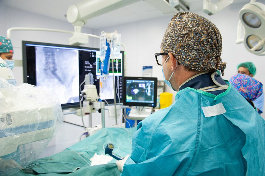
- Venography with intravascular ultrasound (IVUS): a catheter with an ultrasound probe at its tip that measures the compression from the inside. The VIDIO trial showed that IVUS detects and quantifies these lesions better than venography alone and also guides the exact size and placement of the stent.
Treatment
It depends on the syndrome, the severity of the symptoms and whether there has been thrombosis. Many patients improve with conservative measures. When the compression is significant and explains the symptoms, treatment is usually endovascular: through a puncture in the groin, a stent is placed to keep the vein open. In other cases surgery or a combined approach is required. Each indication is decided individually.
Frequently asked questions
Which specialist treats vascular compression syndromes?
A vascular surgeon, who assesses the impact of the compression on blood flow and performs the endovascular or surgical treatments. Depending on the case, we work together with gastroenterology, urology, gynaecology, rehabilitation or general surgery.
Does a compression on a CT scan mean I have the syndrome?
No. These compressions are common in people without symptoms. We speak of a syndrome only when the compression alters blood flow and explains the symptoms.
Do you see patients from outside Mallorca?
Yes. You can send me your test results: I review them in advance and we hold a first video consultation. Here is how it works.
References
- Kibbe MR, Ujiki M, Goodwin AL, Eskandari M, Yao J, Matsumura J. Iliac vein compression in an asymptomatic patient population. J Vasc Surg. 2004;39(5):937-943.
- Gagne PJ, Tahara RW, Fastabend CP, et al. Venography versus intravascular ultrasound for diagnosing and treating iliofemoral vein obstruction. J Vasc Surg Venous Lymphat Disord. 2017;5(5):678-687.
- De Maeseneer MG, Kakkos SK, Aherne T, et al. European Society for Vascular Surgery (ESVS) 2022 Clinical Practice Guidelines on the Management of Chronic Venous Disease of the Lower Limbs. Eur J Vasc Endovasc Surg. 2022;63(2):184-267.
- Ananthan K, Onida S, Davies AH. Nutcracker syndrome: an update on current diagnostic criteria and management guidelines. Eur J Vasc Endovasc Surg. 2017;53(6):886-894.
- Illig KA, Donahue D, Duncan A, et al. Reporting standards of the Society for Vascular Surgery for thoracic outlet syndrome. J Vasc Surg. 2016;64(3):e23-e35.
Patient information reviewed by Dr Óscar Merino, vascular surgeon. It does not replace an individual medical assessment.
Would you like an assessment?
Consultation with duplex ultrasound at Clínica Rotger, in the centre of Palma.
Related
Follow me
Weekly tips on circulation and vascular health (in Spanish).